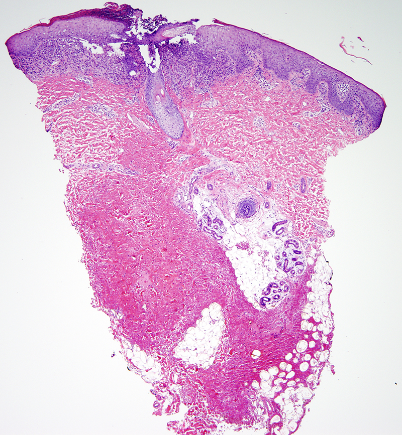
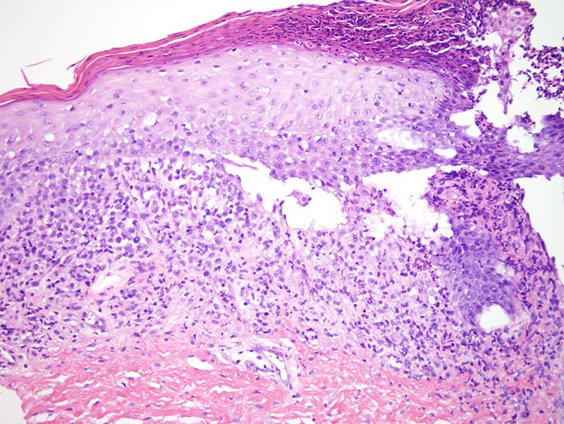
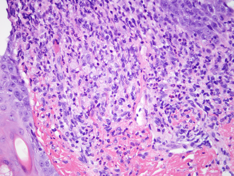
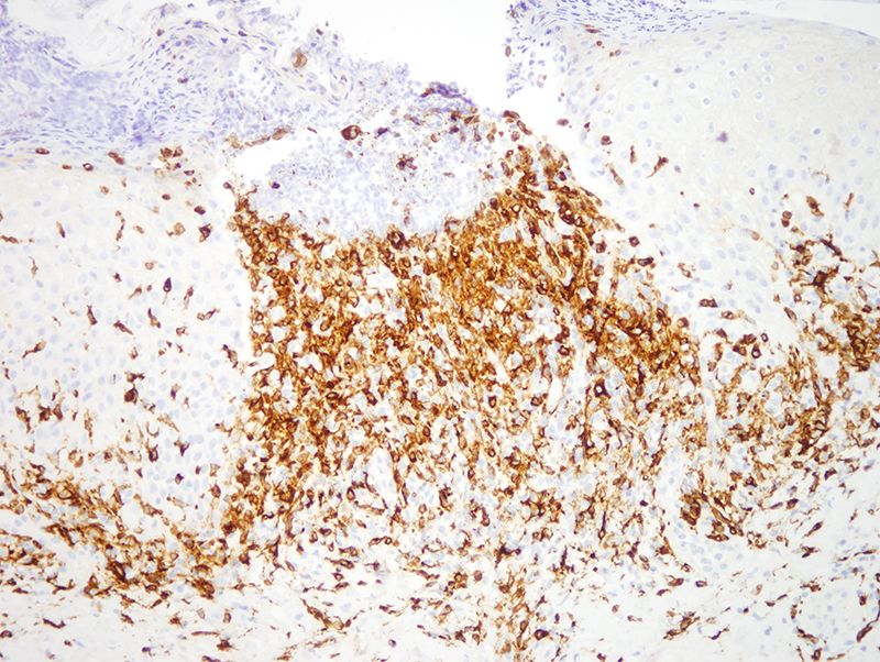
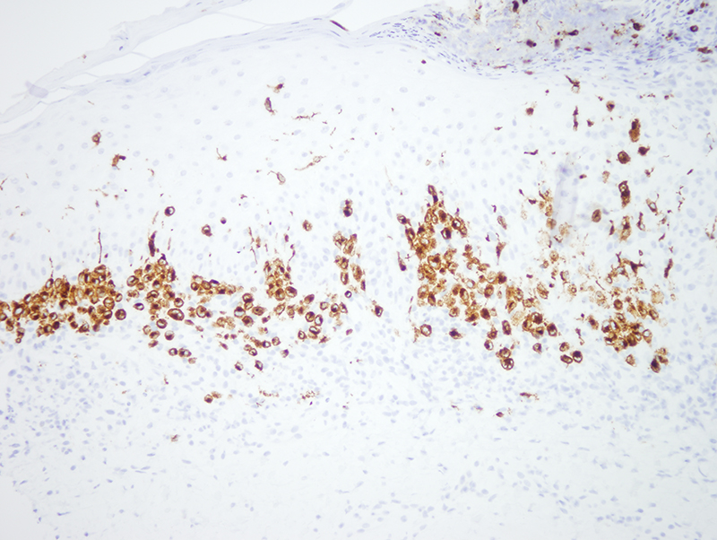
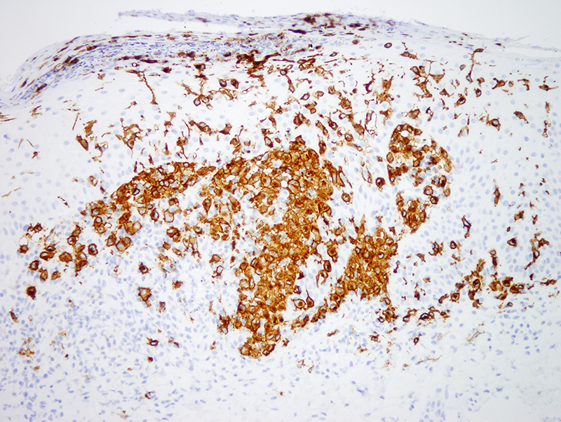
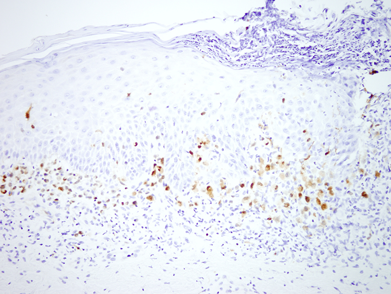
Langerhans cell histiocytosis is characterized by a proliferation of CD1a, langerin (CD207), S100 positive cells presenting as a localized, multifocal, or disseminated disease. Multisystem disease portends a more aggressive clinical course compared to localized disease. The Birbeck granule is seen on electron microscopic examination. The implicated cell typically shows a folded grooved nucleus likened to a coffee bean and is frequently seen in association with eosinophils and scattered lymphocytes.
Click images to begin the slideshow