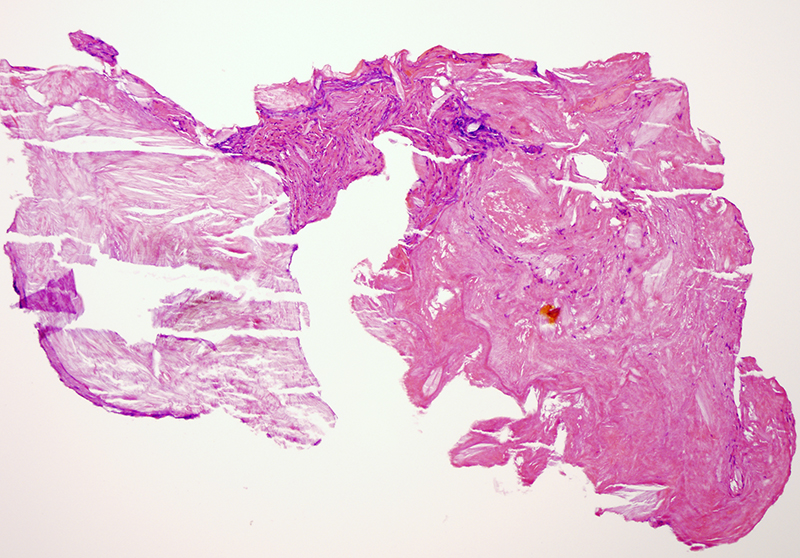
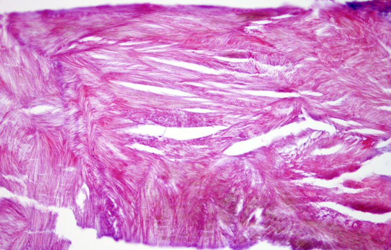
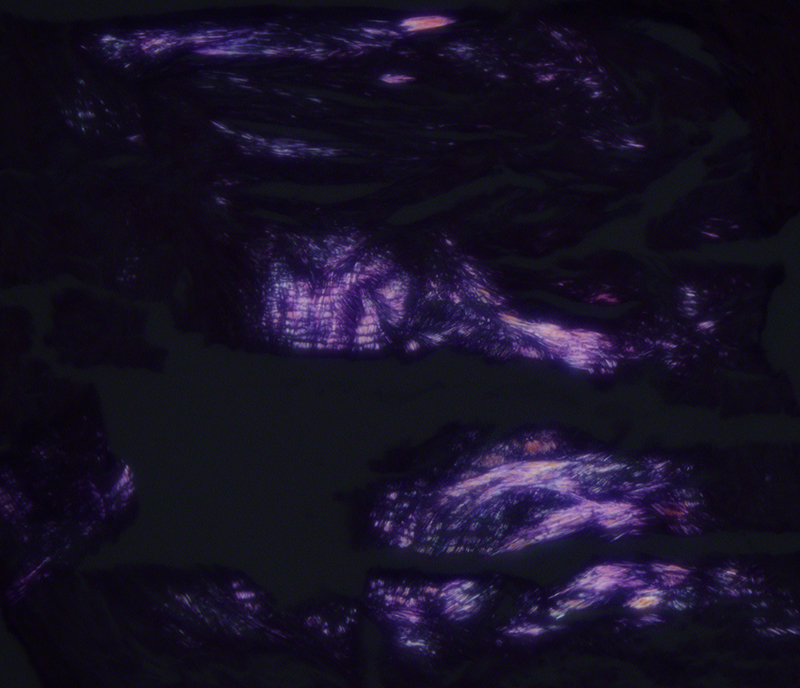
Monosodium urate deposition disease/gout is characterized by extracellular fluid deposits that range from tiny to massive, typically assuming a dermal or subcutaneous location. The clinical presentation can include tophaceous deposits, uric acid kidney stones, recurrent inflammatory arthritis, chronic arthritis or chronic nephropathy. Fixation in alcohol preserves the sodium urate monohydrate crystals. The crystals appear needle-shaped and demonstrate negative birefringence when polarized. Formalin fixation is avoided if one wishes to see the crystals because formalin dissolves them and the deposits appear as amorphous pink areas. While not seen in this case, gouty tophi are frequently associated with a foreign body type giant cell reaction.


